 Reading anthropologist Doug Ubelaker’s recent click! commentary about how photography has been used in the practice of forensic anthropology, especially in the analysis of evidence, brought to mind the photographs, most of them portraits, made by photographer William Bell in the years just after the Civil War. Like the anthropological images of bones and objects left over from human activity, Bell’s images of wounded soldiers constitute an archive where the interesting questions are about what you can see if you know how to look. William Bell was first a soldier, serving in the war between the United States and Mexico in 1846-48, and then a photographer, opening a studio in Philadelphia in 1860. He served in the American Civil War with the First Regiment of Pennsylvania Volunteers. After the war, he became head of the photographic department for the Army Medical Museum.
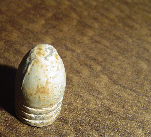
  The original Army Medical Museum (now the National Museum of Health and Medicine) was founded as a research facility in 1862 and collected and commissioned photographs to study specimens of morbid anatomy, surgical or medical. At the beginning of the Civil War, newly enlisted doctors had almost no experience with gunshot wounds, especially those made by the recently developed Minié ball. Shaped like a pointed cone, this high-speed bullet caused significantly worse wounds than the older lead balls. In an era before X-Rays, short of dissecting the body, no one knew what a wound looked like inside of damaged tissue. Bell and the photographers who succeeded him at the Army Medical Museum carefully documented the effects rather than the events of the war.
 The seven-volume Photographic Catalogue of the Surgical Section of the Army Medical Museum, begun in 1865, included detailed case histories and fifty tipped-in albumen prints. Photographs of shattered bones and skulls display an appropriately clinical approach to the subject of scientific inquiry. The portraits of the wounds of survivors, however, command (and are arguably compromised by) a more emotional scrutiny. These elegant, studio-style portraits are unnervingly intimate. Formal science is linked with artistic formality. Along with the catalogue’s detailed descriptions of the affliction and the appropriate medical procedure, the photographs were useful to doctors who wondered just what the slice or dice they contemplated might look like when finished. Today, it is hard to know where to cast your eye in these pictures. The subjects (it is hard to refer to them in the vocabulary of portraiture as “sitters”) often gaze intensely, and considering the extent of both their disfigurement and state of undress, unabashedly, at the camera. I wonder, is it the face or the wound that gives us the most information about war? Bell’s photographs of mutilated soldiers suggest the near-impossibility of simultaneous looking and seeing. These images of wounds and wounded still are so very beautiful, and heartbreaking. See more medical photographs by Bell from the Smithsonian American Art Museum here. For a more complete description of surgical practice during the Civil War go to the National Museum of Health and Medicine’s on line exhibition, Trauma and Surgery: Medicine During the Civil War.
Merry Foresta is the Former Director of the Smithsonian Photography Initiative.
Produced by the Smithsonian Institution Archives. For copyright questions, please see the Terms of Use.

Leave a Comment